Corneal Imaging and Other Technology at Paul Vision Institute
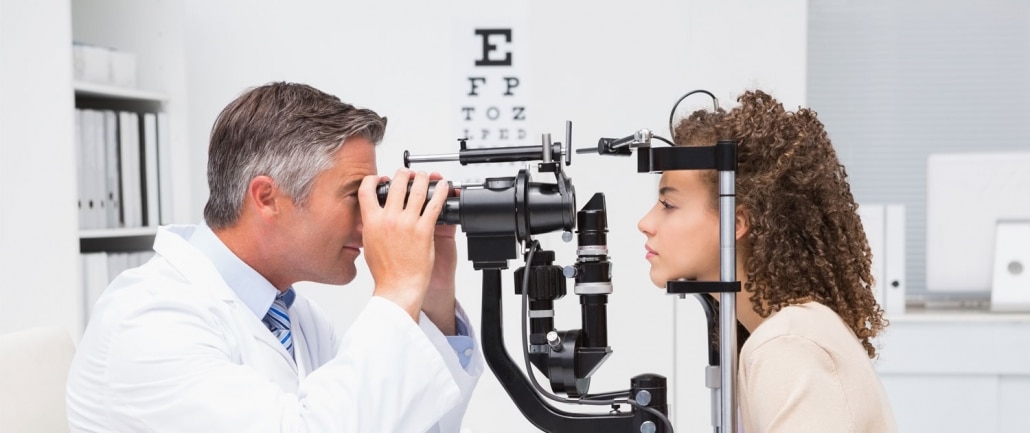
Corneal mapping in Wilmington, NC, is a technology that goes by many different names. You may also hear Dr. Paul or another staff member at Paul Vision Institute refer to it as corneal topography, video keratography, or photokeratoscopy. Dr. Edward Paul invested in this technology to enable our optometrists to map out the curvature of the cornea in both eyes.
What is the Cornea?
Your corneas sit in the outermost layer of your eyes and cover the anterior chamber, iris, and pupil. The primary function of the cornea is to refract light as it enters your eyes, allowing you to interpret images correctly. Another job of the cornea is to protect your eyes from dirt, grit, germs, and any other potentially harmful substances that could become trapped in them.
How Does Corneal Mapping Work?
This painless procedure sends color-coded images to a computer screen to assist our optometrists in diagnosing various visual conditions. Mapping can help the optometrist working with you pick up on even subtle changes in your vision and/or eye health. Some of the specific things that our optometrists measure with mapping include the thickness of your corneas and any elevation changes.
Corneal mapping is especially useful for helping to plan eye surgery, such as LASIK or cataracts, and diagnosing the eye conditions listed below.
- Astigmatism
- Keratoconus, which is an optical condition that causes your corneas to take on a cone-shaped appearance and dulls your vision
- Scarring from previous infections or injuries
- Unusual growths
If you are concerned that your vision is becoming less sharp or you have sustained trauma to your eyes in the past, please do not hesitate to ask us about corneal mapping in Wilmington, NC. All you have to do is mention your symptoms or that you would like to have the test when scheduling your next eye exam.